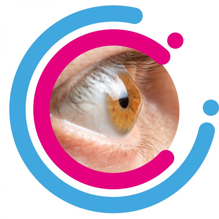
Keratoconus
Keratoconus is the most common dystrophy of the cornea, affecting around one person in a thousand, although some reports indicate prevalence as high as 1 in 500 individuals. It is typically diagnosed in the mid to late teens and attains its most severe state in the twenties and thirties.

Keratoconus in detail
Keratoconus is a degenerative non-inflammatory disorder of the cornea (the front window of the eye) and generally affects both eyes. The underlying problem is weakness of the supporting collagen fibres in the cornea. This makes the cornea structurally and biomechanically “weak”. As a result, the cornea assumes a more conical shape with resultant irregular astigmatism.
The progression of Keratoconus can be quite variable, with some patients remaining stable while others progress rapidly or experience occasional exacerbations over a long and otherwise steady course. A genetic predisposition to keratoconus has been observed, with the disease running within families in 10% of all cases.
How is Keratoconus diagnosed?
From the patient’s history, a detailed slit lamp examination as well as sophisticated corneal imagery which can assess the profile of the cornea.
Symptoms of Keratoconus
Symptoms can include substantial distortion of vision (astigmatism), with multiple images, blurry (near and far sighted) vision and sensitivity to light (photophobia). Initially most people can correct their vision with glasses. But as the astigmatism worsens, most patients can be managed with specially fitted rigid gas permeable contact lenses to reduce the distortion and provide better vision. Symptoms may be unilateral initially and may later become bilateral. In 20% of patients, the condition is progressive and requires surgical intervention.

What are the surgical interventions?
Corneal INTACS
Corneal Collagen Crosslinking
Corneal Transplantation
Get in touch
Please contact us if you have any queries or questions about our eye examination and related issues, we would be most happy to advise you.
